Dark-field Microscopy Depends on Which Characteristic of Light
Hexagonal polytype phase flakes. Wall cannot be seen by direct light microscopy and does not stain with simple.

Principle Of Dark Field Microscopy Download Scientific Diagram
Due to its many protean clinical manifestations it has been named the great imitator and mimicker The origin of syphilis has been controversial and under great debate and many theories have been postulated regarding this.
. Treponema pallidum subsp pallidum exhibits characteristic motility that consists of rapid rotation about its longitudinal axis and bending flexing and snapping about its full length. He knew that electrons possess a wave aspect so he believed he could treat them in a fashion similar to light waves. Dark field microscope c light beam.
Using high-angle annular dark-field imaging in scanning transmission electron microscopy HAADF-STEM we studied the atomic configuration of the ML 2H-WSe 2 ie. It is a specific case of electron diffraction used primarily in material science and solid state physics as one of the most common experimental techniques. Optical or light microscopy involves passing visible light transmitted through or reflected from the sample through a single lens or multiple lenses to allow a magnified view of the sample.
Transmission Electron Microscopy-TEM-The first electron microscope was built 1932 by the German physicist Ernst Ruska who was awarded the Nobel Prize in 1986 for its invention. Compared with other types of microscopy for surface imaging such as SEM and TEM optical microscopy is restricted by the diffraction limit of visible light to 1000 magnification. Numerous nanophotonic biosensors have.
Especially with appropriate analytical. Live treponemes which are too slender to be seen by conventional light microscopy can be visualized by using dark-field microscopy. To overcome this fluorescence dark field and phase-contrast optical microscopy techniques have been developed leading to microscopic images with sufficient contrast and high information content.
Syphilis is a systemic bacterial infection caused by the spirochete Treponema pallidum. Production depends on growth conditions. Selected area electron diffraction abbreviated as SAD or SAED is a crystallographic experimental technique typically performed using a transmission electron microscope TEM.
Enter the email address you signed up with and well email you a reset link. The resulting image can be detected directly by the eye imaged on a photographic plate or captured digitallyThe single lens with its attachments or the system of lenses and imaging equipment. Nanophotonic devices which control light in subwavelength volumes and enhance lightmatter interactions have opened up exciting prospects for biosensing.
This nice numerical analysis to study differential equation.
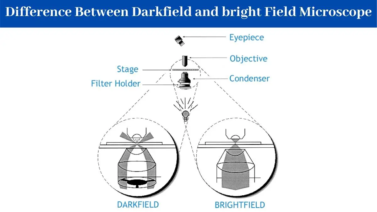
12 Difference Between Darkfield And Bright Field Microscope

What Is A Dark Field Microscopy And How Is It Advantageous Compared To The Bright Field Or Phase Contrast Microscopy Quora

What Is A Dark Field Microscopy And How Is It Advantageous Compared To The Bright Field Or Phase Contrast Microscopy Quora
No comments for "Dark-field Microscopy Depends on Which Characteristic of Light"
Post a Comment